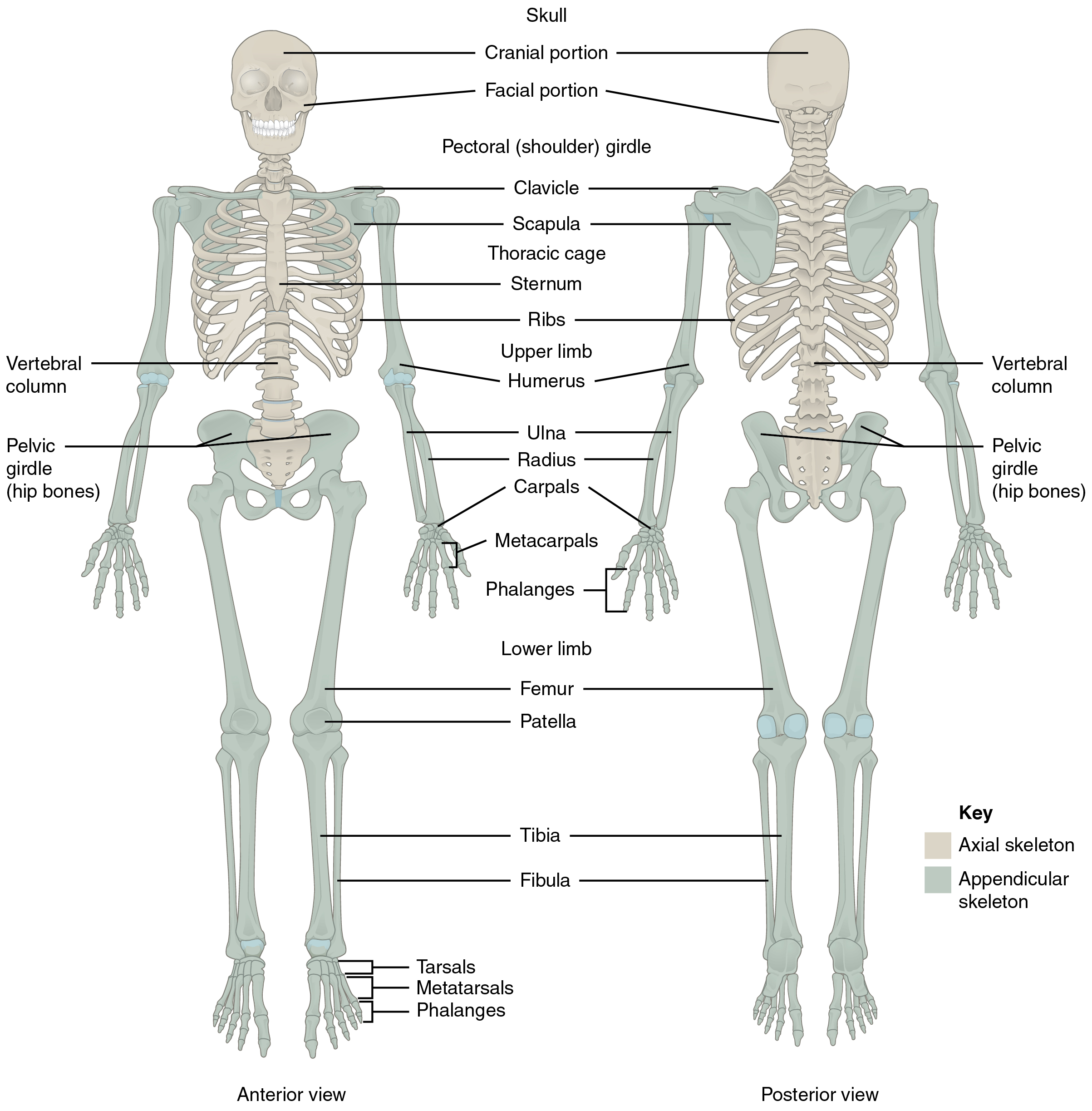
 anterior and posterior views of the skeleton. Image description available." width="500" height="2447" />
Click on prefixes, combining forms, and suffixes to reveal a list of word parts to memorize for the Skeletal System.
The skeletal system forms the framework of the body. It is the body system composed of bones, cartilage, and ligaments. Each bone serves a particular function and varies in size, shape, and strength. Bones are weight-bearing structures in your body and can therefore change in thickness as you gain or lose weight. The skeletal system performs the following critical functions for the human body:
The skeletal system includes all of the bones, cartilages, and ligaments of the body that support and give shape to the body and body structures. The skeleton consists of the bones of the body. For adults, there are 206 bones in the skeleton. Younger individuals have higher numbers of bones because some bones fuse together during childhood and adolescence to form an adult bone. The primary functions of the skeleton are to provide a rigid, internal structure that can support the weight of the body against the force of gravity, and to provide a structure upon which muscles can act to produce movements of the body.
In addition to providing for support and movements of the body, the skeleton has protective and storage functions. It protects the internal organs, including the brain, spinal cord, heart, lungs, and pelvic organs. The bones of the skeleton serve as the primary storage site for important minerals such as calcium and phosphate. The bone marrow found within bones stores fat and houses the blood-cell-producing tissue of the body.
The skeleton is subdivided into two major divisions: the axial and appendicular.
The axial skeleton forms the vertical, central axis of the body and includes all bones of the head, neck, chest, and back (see Figure 6.1). It serves to protect the brain, spinal cord, heart, and lungs. It also serves as the attachment site for muscles that move the head, neck, and back, and for muscles that act across the shoulder and hip joints to move their corresponding limbs.
The axial skeleton of the adult consists of 80 bones, including the skull, the vertebral column, and the thoracic cage. The skull is formed by 22 bones. Also associated with the head are an additional seven bones, including the hyoid bone and the ear ossicles (three small bones found in each middle ear). The vertebral column consists of 24 bones, each called a vertebra, plus the sacrum and coccyx. The thoracic cage includes the 12 pairs of ribs and the sternum, the flattened bone of the anterior chest.
anterior and posterior views of the skeleton. Image description available." width="500" height="2447" />
The cranium or skull supports the face and protects the brain. It is subdivided into the bones of the skull and the bones of the face.
The axial skeleton has 80 bones and includes bones of the s kull (and face), v ertebral column, and t horacic cage.
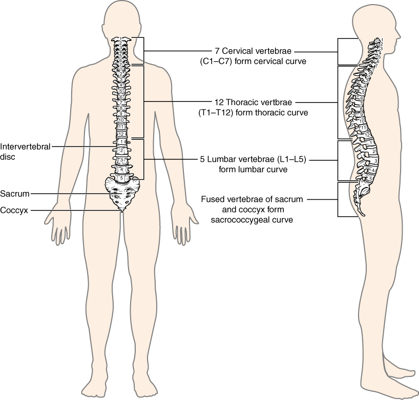
The vertebral column is also known as the spinal column or spine (see Figure 6.2). It consists of a sequence of vertebrae (singular = vertebra), each of which is separated and united by an intervertebral disc. Together, the vertebrae and intervertebral discs form the vertebral column. It is a flexible column that supports the head, neck, and body and allows for their movements. It also protects the spinal cord, which passes down the back through openings in the vertebrae.
The thoracic cage (rib cage) forms the thorax (chest) portion of the body. It consists of the 12 pairs of ribs with their costal cartilages and the sternum (see Figure 6.3). The ribs are anchored posteriorly to the 12 thoracic vertebrae (T1–T12). The thoracic cage protects the heart and lungs.
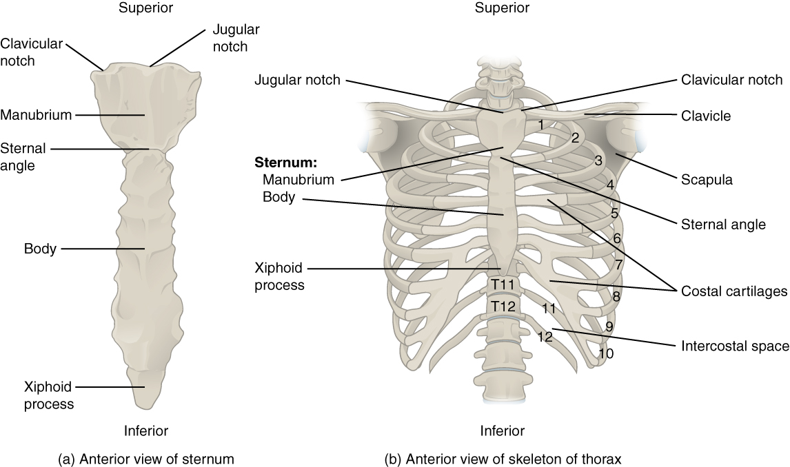
There are 12 sets of ribs and can be divided as such:
The sternum, also known as the breast bone, is divided into 3 parts:
The appendicular skeleton includes all bones of the upper and lower limbs, plus the bones that attach each limb to the axial skeleton. There are 126 bones in the appendicular skeleton of an adult.
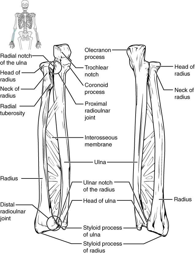
The bones of the upper limbs include the bones of the arms, wrists, and hands.
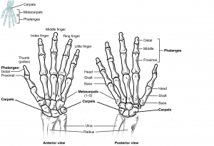
Each phalanx has three bones: the distal, medial, and proximal. The exception is the thumb and big toe which has two bones: the distal and proximal (Figure 6.5). There are 30 bones in each upper limb. Can you count them on your limb?
The appendicular skeleton has 126 bones. It is divided into the bones of the u pper limbs and l ower limbs that attach each limb to the skeleton.
The bones of the pelvic region protect the reproductive, urinary, and excretory organs.
The shape of the pelvic girdle is different for males than females. In the male, it is a funnel shape. In the female, it is shaped like a basin to accommodate the fetus during pregnancy.
The bones of the lower limb include bones of the leg and the feet.
The femur is the longest and strongest bone of the body and accounts for approximately one-quarter of a person’s total height.
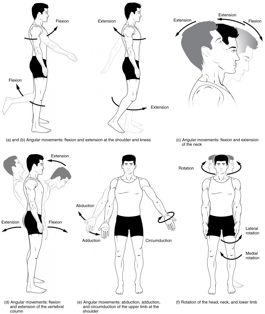
Flexion and extension are movements that take place within the sagittal plane and involve anterior or posterior movements of the body or limbs. For the vertebral column, flexion (anterior flexion) is an anterior (forward) bending of the neck or body, while extension involves a posterior-directed motion, such as straightening from a flexed position or bending backward. Lateral flexion is the bending of the neck or body toward the right or left side. These movements of the vertebral column involve both the joints as well as the associated intervertebral disc.
In the limbs, flexion decreases the angle between the bones (bending of the joint), while extension increases the angle and straightens the joint (see Figures 6.8(a-d)). You will discover in the muscular system chapter that the associated muscles to these movements are flexor and extensor.
Abduction and adduction motions occur within the coronal plane and involve medial-lateral motions of the limbs, fingers, toes, or thumb. For example, abduction is raising the arm at the shoulder joint, moving it laterally away from the body, while adduction brings the arm down to the side of the body (see Figure 6.8(e)). In the muscular system chapter, you will discover that the associated muscles to these movements are the abductor and adductor.
Circumduction is the movement of a body region in a circular manner, in which one end of the body region being moved stays relatively stationary while the other end describes a circle. It involves the sequential combination of flexion, adduction, extension, and abduction at a joint (see Figure 6.8(e)).
Rotation can occur within the vertebral column, at a pivot joint, or at a ball-and-socket joint. Rotation of the neck or body is the twisting movement produced by the summation of the small rotational movements available between adjacent vertebrae. At a pivot joint, one bone rotates in relation to another bone.
Rotation can also occur at the ball-and-socket joints of the shoulder and hip. Here, the humerus and femur rotate around their long axis, which moves the anterior surface of the arm or thigh either toward or away from the midline of the body (see Figure 6.8(f)).
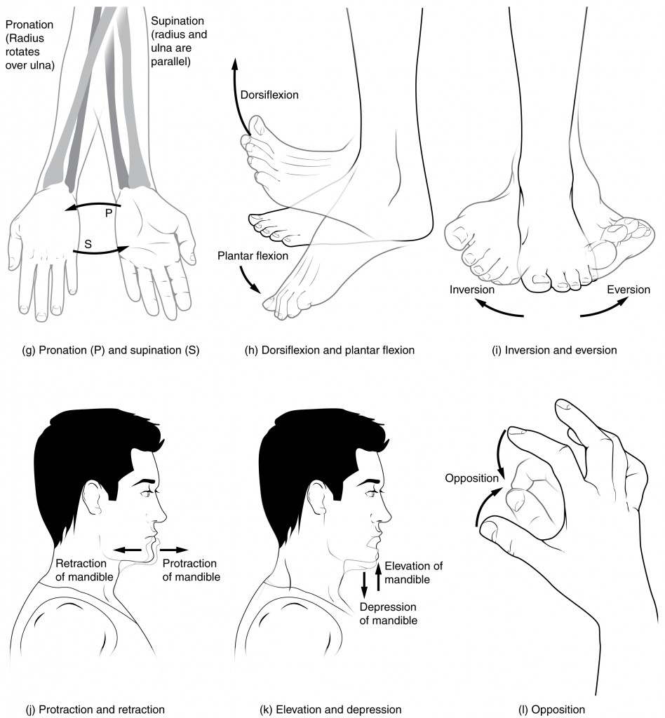
Supination and pronation are movements of the forearm. In the anatomical position, the upper limb is held next to the body with the palm facing forward. This is the supinated position of the forearm. In this position, the radius and ulna are parallel to each other. When the palm faces backward, the forearm is in the pronated position, and the radius and ulna form an X-shape.
Pronation is the movement that allows the palm to face backward while in supination the palm faces forward. It helps to remember that supination is the motion you use when scooping up soup with a spoon (see Figure 6.9(g)).
Dorsiflexion and plantar flexion are movements at the ankle joint, which is a hinge joint. Lifting the front of the foot, so that the top of the foot moves (upward) toward the anterior leg is dorsiflexion, while lifting the heel of the foot from the ground or pointing the toes downward is plantar flexion. These are the only movements available at the ankle joint (see Figure 6.9(h)).
Inversion and eversion are complex movements that involve the multiple plane joints among the tarsal bones of the posterior foot (intertarsal joints) and thus are not motions that take place at the ankle joint. Inversion is the turning of the foot to angle the bottom of the foot toward the midline, while eversion turns the bottom of the foot away from the midline. The foot has a greater range of inversion than eversion motion. These are important motions that help to stabilize the foot when walking or running on an uneven surface and aid in the quick side-to-side changes in direction used during active sports such as basketball, racquetball, or soccer (see Figure 6.9(i)).
Protraction and retraction are anterior-posterior movements of the scapula or mandible. Protraction of the scapula occurs when the shoulder is moved forward, as when pushing against something or throwing a ball. Retraction is the opposite motion, with the scapula being pulled posteriorly and medially, toward the vertebral column. For the mandible, protraction occurs when the lower jaw is pushed forward, to stick out the chin, while retraction pulls the lower jaw backward (see Figure 6.9(j)).
Depression and elevation are downward and upward movements of the scapula or mandible. The upward movement of the scapula and shoulder is elevation, while a downward movement is depression. These movements are used to shrug your shoulders. Similarly, elevation of the mandible is the upward movement of the lower jaw used to close the mouth or bite on something, and depression is the downward movement that produces the opening of the mouth (see Figure 6.9(k)).
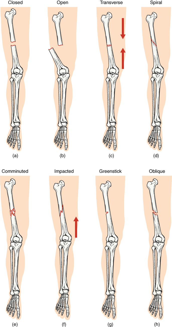
A fracture is a broken bone. It will heal whether or not a physician resets it in its anatomical position. If the bone is not reset correctly, the healing process will keep the bone in its deformed position. Crepitation or crepitus is the creaking or popping sound that is heard when fractured bones move against each other. Fractures are classified by their complexity, location, and other features (see Figure 6.12). Some fractures may be described using more than one term because they may have the features of more than one type (e.g., an open transverse fracture).
Types of fractures include:
There are three types of primary bone cancers: osteosarcoma, Ewing Sarcomas, and chondrosarcoma. These are considered primary cancers because they originate in the bones. Osteosarcoma and Ewing Sarcomas primarily affect children, teenagers, and young adults. Chondrosarcoma primarily affects older adults (National Cancer Institute, n.d.-a). To learn more, visit the American Cancer Society’s web page on bone cancer.
Orthopedic surgeons are medical doctors who have specialized training in the prevention, diagnosis, treatment, and surgery of disorders and diseases related to the musculoskeletal systems (Bureau of Labor Statistics, 2021a). For more details, please visit the American College of Surgeons’ page on Orthopedic Surgery.
Rheumatologists are medical doctors who specialize in the diagnosis and treatment of disorders of the joints, muscles, and bones. They diagnose and treat diseases such as arthritis, musculoskeletal disorders, osteoporosis, plus autoimmune diseases like ankylosing spondylitis, a chronic spinal inflammatory disease, and rheumatoid arthritis (Fowler et al., 2013). For more details, please follow the link to the American College of Rheumatology’s page on rheumatology.
Chiropractors are required to have a Doctor of Chiropractic (D.C.) degree, which is a 4-year postgraduate professional degree, and a state license. Chiropractors focus on spinal adjustments, nutrition, and preventing injury without the use of pharmaceuticals or surgical procedures (Bureau of Labor Statistics, 2021b). To learn more, visit the Bureau of Labor Statistics’ website.
A physical therapist is a licensed professional who develops individualized treatment plans for their clients. These plans can include exercises, hands-on therapy, and equipment, such as canes or wheelchairs. Although current licensure laws require that those entering the field have a doctor of physical therapy degree, physical therapists who began working before those requirements went into effect may have a bachelor’s or master’s degree (Bureau of Labor Statistics, 2021c). To learn more, please visit the American Physical Therapy Association website.
Common diagnostic procedures related specifically to the skeletal system include x-rays, bone mineral density testing, and arthroscopy.
Abduction
Moving the limb or hand laterally away from the body, or spreading the fingers or toes.
Adduction
Movement that brings the limb or hand toward or across the midline of the body, or brings the fingers or toes together.
Amphiarthrosis
A slightly mobile joint.
Ankylosis
Fixation and immobility of a joint.
Appendicular skeleton
All bones of the upper and lower limbs, plus the girdle bones that attach each limb to the axial skeleton.
Arthralgia
Arthritis
Chronic inflammation of the synovial joints.
Arthrocentesis
Surgical puncture to aspirate fluid from a joint.
Arthrodesis
Surgical fixation of a joint.
Arthrography
Process of recording a joint.
Arthroplasty
Joint replacement surgery.
Arthroscopy
Process of viewing a joint using an endoscope.
Articulations
Where two bone surfaces meet.
Autoimmune diseases/disorders
Disorders in which the immune system overreacts and begins to attack itself.
Axial skeleton
The central, vertical axis of the body, including the skull, vertebral column, and thoracic cage.
Bradykinesia
Condition of slow movement.
Bursitis
Inflammation of a bursa near a joint.
Chondromalacia
Degeneration of cartilage.
Chronic
A condition that lasts a long time with periods of remission and exacerbation.
Craniotomy
An operation in which a piece of the skull is removed.
Diarthrosis
Freely mobile joints.
Diskectomy
Excision of the intervertebral disk.
Inflammation of the intervertebral disk.
Dyskinesia
Abnormal involuntary movements of the extremities, trunk, or jaw.
Edema
Swelling due to excessive liquid in the tissues.
Eversion
Foot movement in which the bottom of the foot is turned laterally, away from the midline.
Extension
Movement in the sagittal plane that increases the angle of a joint (straightens the joint).
Flexion
Movement in the sagittal plane that decreases the angle of a joint (bends the joint).
Hematopoiesis
The production of blood cells.
Hyperkinesia
Excessive movement of muscles of the body as a whole.
Hypertrophy
The enlargement of muscles.
Inversion
Foot movement in which the bottom of the foot is turned toward the midline.
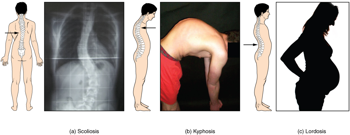
Kyphosis
An excessive posterior curvature of the thoracic region; also called humpback.
Lordosis
Excessive anterior curvature of the lumbar vertebral column region; also called swayback.
Lumbar
Pertaining to the lumbar region of the spine (L1 to L5).
Lumbosacral
Pertaining to the region of the back that includes the lumbar vertebrae, sacrum, and nearby structures.
Muscular dystrophy
A general term for the group of inherited myopathies that are characterized by wasting and weakness of the skeletal muscle.
Osteitis
Osteoarthritis
The most common type of arthritis; associated with aging and “wear and tear” of the articular cartilage.
Osteoblast
The cell responsible for forming new bone.
Osteochondritis
Inflammation of bone and cartilage.
Osteocyte
Osteomalacia
A softening of adult bones due to Vitamin D deficiency.
Osteomyelitis
Inflammation of bone and bone marrow.
Osteonecrosis
Abnormal condition of bone death (lack of blood supply).
Osteopenia
Abnormally low bone mass or bone mineral density.
Osteopetrosis
Abnormal condition of porous bones.
Osteoporosis
A disease characterized by a decrease in bone mass that occurs when the rate of bone resorption exceeds the rate of bone formation.
Osteosarcoma
Malignant tumor of bone.
Pelvic
Pertaining to the pelvis.
Pronation
Forearm motion that moves the palm of the hand from the palm forward to the palm backward position.
Rotation
Movement of a bone around a central axis or around its long axis.
Sarcopenia
Age-related muscle atrophy.
Scoliosis
Lateral curvature of the spine.
Spondyloarthritis
Inflammation of the joints of the spine.
Spondylosis
A degenerative spinal disease that can involve any part of the vertebra, intervertebral disk, and surrounding soft tissue.
Supination
Forearm motion that moves the palm of the hand from the palm backward to the palm forward position.
Synarthrosis
An immobile or nearly immobile joint.
Synovectomy
Excision of the synovial membrane.
Synovial sarcoma
Malignant tumor of the synovial membrane.
Tendinitis
Inflammation of the tendon.
Tenosynovitis
Inflammation of the synovial membrane of a tendon.
Vertebroplasty
A procedure used to repair a bone in the spine that has a break caused by cancer, osteoporosis, or trauma.
Bureau of Labor Statistics. (2021a). Physicians and surgeons. In Occupational outlook handbook. U.S. Department of Labor. https://www.bls.gov/ooh/healthcare/physicians-and-surgeons.htm
Bureau of Labor Statistics. (2021b). Chiropractors. In Occupational outlook handbook. U.S. Department of Labor. https://www.bls.gov/ooh/healthcare/chiropractors.htm
Bureau of Labor Statistics. (2021c). Physical therapists. In Occupational outlook handbook. U.S. Department of Labor. https://www.bls.gov/ooh/healthcare/physical-therapists.htm
Centers for Disease Control and Prevention. (n.d.-b). Rheumatoid arthritis. CDC Arthritis Program. https://www.cdc.gov/arthritis/basics/rheumatoid-arthritis.html
MedlinePlus. (2021). X-rays. U.S. National Library of Medicine. https://medlineplus.gov/xrays.html
National Cancer Institute. (n.d.-a). Primary bone cancer [Fact sheet]. National Institutes of Health, U.S. Department of Health and Human Services. https://www.cancer.gov/types/bone/bone-fact-sheet
National Cancer Institute. (n.d.-b). Dual x-ray absorptiometry. National Institutes of Health, U.S. Department of Health and Human Services. https://www.cancer.gov/publications/dictionaries/cancer-terms/def/dual-x-ray-absorptiometry
National Institute of Arthritis and Musculoskeletal and Skin Diseases. (n.d.-a). Arthritis. National Institutes of Health, U.S. Department of Health and Human Services. https://www.niams.nih.gov/health-topics/arthritis
National Institute of Arthritis and Musculoskeletal and Skin Diseases. (n.d.-b). Osteoporosis overview. National Institutes of Health, U.S. Department of Health and Human Services. https://www.bones.nih.gov/health-info/bone/osteoporosis/overview
National Institutes of Arthritis and Musculoskeletal and Skin Diseases. (n.d-c). Health topics: Fibromyalgia [PDF]. National Institutes of Health, U.S. Department of Health and Human Services. https://www.niams.nih.gov/print/view/pdf/advanced_reading_pdf_/advanced?view_args%5B0%5D=1957
Office of Communications and Public Liaison. (2020). Myasthenia gravis fact sheet. National Institute of Neurological Disorders and Stroke, U.S. Department of Health and Human Services. https://www.ninds.nih.gov/Disorders/Patient-Caregiver-Education/Fact-Sheets/Myasthenia-Gravis-Fact-Sheet
Figure 6.1 image description: This diagram shows the human skeleton and identifies the major bones. The left panel shows the anterior view (from the front) and the right panel shows the posterior view (from the back). Labels read (from the top of the skull): skull (cranial portion, facial portion), pectoral shoulder girdle, clavicle, scapula, thoracic cage (sternum, ribs), upper limb (humerus, ulna, radius, carpals, metacarpals, phalanges), vertebral column, pelvic girdle (hip bones), lower limb (femur, patella, tibia, fibula, tarsals, metatarsals, phalanges). [Return to Figure 6.1].
Figure 6.2 image description: This image shows the structure of the vertebral column. The left panel shows the front view of the vertebral column. Labels and the right panel show the side view of the vertebral column. labels read (from top): 7 cervical vertebrae (C1-C7) form cervical curve, 12 thoracic vertebrae (T1-T12) form the thoracic curve, intervertebral disc, 5 lumbar vertebrae (L1-L5) form lumbar curve, Fused vertebrae of sacrum and coccyx form a sacrococcygeal curve, sacrum, coccyx. [Return to Figure 6.2].
Figure 6.3 image description: This figure shows the skeletal structure of the rib cage. The left panel shows the anterior view of the sternum. Labels read (from top): clavicular notch, jugular notch, manubrium, sternal angle, body, xiphoid process. The right panel shows the anterior panel of the sternum including the entire rib cage. Labels read (from top): jugular notch, clavicular notch, clavicle, sternum (manubrium, body, xiphoid process), scapula, sternal angle, costal cartilages, intercostal space. Ribs are numbered 1-12 from the top. [Return to Figure 6.3].
Figure 6.4 image description: This diagram labels the bones of the lower arm (excluding the hands). Labels read (from top): olecranon process, head of radius, radial notch of the ulna, trochlear notch, coronoid process, radial tuberosity, proximal radioulnar joint, neck of radius, radius, interosseous membrane, ulna, ulnar notch of the radius, head of the ulna, distal radioulnar joint, styloid process of ulna, styloid process of radius. [Return to Figure 6.4].
Figure 6.5 image description: This diagram shows an anterior and posterior view of the hands with corresponding labels. Anterior view labels read (from top): middle finger, ring finger, index finger, little finger, thumb, phalanges (distal, proximal), metacarpals, carpals, ulna, radius. Posterior view labels read (from top): Phalanges (distal, middle, proximal), head shaft and base of the proximal phalanx, head shaft and base of the metatarsal, metatarsals 1-5, carpals, ulna, radius. [Return to Figure 6.5].
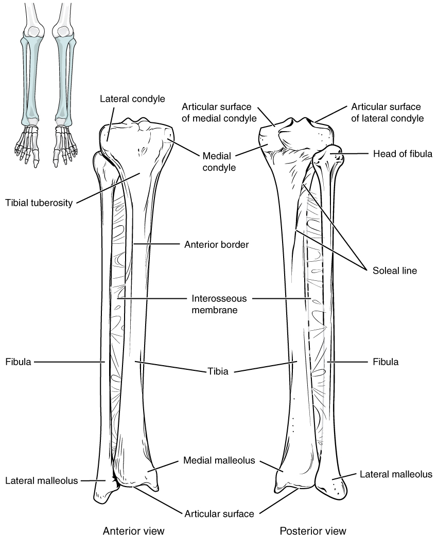
Figure 6.6 image description: This image shows the structure of the tibia and the fibula. The left panel shows the anterior view. Labels read (from top): lateral condyle, medial condyle, tibial tuberosity, anterior border, interosseous membrane, fibula, tibia, medial malleolus, lateral malleolus, articular surface. The right panel shows the posterior view. Labels read (from top): the articular surface of medial and lateral condyles, medial condyle, head of the fibula, soleal line, interosseous membrane, tibia, fibula, medial malleolus, lateral malleolus, articular surface. [Return to Figure 6.6].
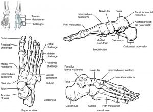
Figure 6.7 image description: This figure shows the bones of the foot. The left panel shows the superior view. Labels read (from toes): distal, proximal phalanges, distal phalange, middle phalange, proximal phalanx, medial cuneiform, intermediate and lateral cuneiforms, navicular, cuboid, talus, trochlea of talus, calcaneus. The top right panel shows the medial view. Labels read (from left to right starting at toe): first metatarsal, medial cuneiform, intermediate cuneiform, navicular, talus, calcaneus, facet for medial malleolus, sustentaculum tali (talar shelf), calcaneal tuberosity. The bottom right panel shows the lateral view. Labels read (from left at the heel, to right): calcaneus, talus, facet for lateral malleolus, cuboid, navicular, intermediate and lateral cuneiforms, fifth metatarsal. [Return to Figure 6.7].
Figure 6.8 image description: This multi-part image shows different types of movements that are possible by different joints in the body. Labels read (from the top, left): a and b angular movements: flexion and extension at the shoulders and knees, c) angular movements: flexion and extension of the neck (arrows pointing left and right to indicate movement). Labels (from the bottom, left) read d) angular movements: flexion and extension of the vertical column, e) angular movements abduction, adduction, and circumduction of the upper limb at the shoulder, f) rotation of the head, neck, and lower limb. [Return to Figure 6.8].
Figure 6.9 image description: This multi-part image shows different types of movements that are possible by different joints in the body. The top left image shows a hand and forearm in the pronation and supination positions. The top middle image shows a foot in the dorsiflexion and plantar flexion positions. The top right image shows a foot in the inversion and eversion positions. The bottom left image shows the retraction and protraction of a man’s mandible. The bottom middle image shows the elevation and depression of a man’s mandible. The bottom right image shows a hand in the opposition position. [Return to Figure 6.9].
Figure 6.10 image description: This image shows the changes to the abnormal curves of the vertebral columns in different diseases. The left panel shows the change in the curve of the vertebral column in scoliosis, the middle panel shows the change in the curve of the vertebral column in kyphosis, and the right panel shows the change in the curve of the vertebral column in lordosis. [Return to Figure 6.10].
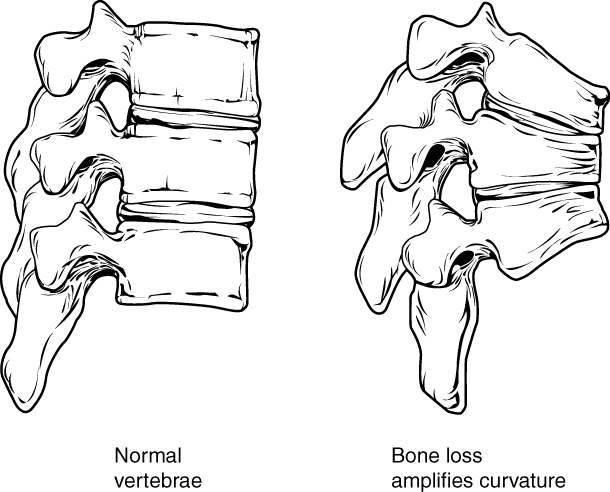
Figure 6.11 image description: This figure shows the changes to the spine in osteoporosis. The left panel shows the structure of normal vertebrae and the right panel shows the curved vertebrae in osteoporosis. [Return to Figure 6.11].
Figure 6.12 image description: In this illustration, each type of fracture is shown on the right femur from an anterior view. In the closed fracture, the femur is broken in the middle of the shaft with the upper and lower halves of the bone completely separated. However, the two halves of the bones are still aligned in that the broken edges are still facing each other. In an open fracture, the femur is broken in the middle of the shaft with the upper and lower halves of the bone completely separated. Unlike the closed fracture, in the open fracture, the two bone halves are misaligned. The lower half is turned laterally and it has protruded through the skin of the thigh. The broken ends no longer line up with each other. In a transverse fracture, the bone has a crack entirely through its width, however, the broken ends are not separated. The crack is perpendicular to the long axis of the bone. Arrows indicate that this is usually caused by compression of the bone in a superior-inferior direction. A spiral fracture travels diagonally through the diameter of the bone. In a comminuted fracture, the bone has several connecting cracks at its middle. The bone could splinter into several small pieces at the site of the comminuted fracture. In an impacted fracture, the crack zig zags throughout the width of the bone like a lightning bolt. An arrow indicates that these are usually caused by an impact that pushes the femur up into the body. A greenstick fracture is a small crack that does not extend through the entire width of the bone. The oblique fracture shown here is traveling diagonally through the shaft of the femur at about a thirty degree angle. [Return to Figure 6.12].
Unless otherwise indicated, this chapter contains material adapted from Anatomy and Physiology (on OpenStax), by Betts et al. and is used under a CC BY 4.0 international license. Download and access this book for free at https://openstax.org/books/anatomy-and-physiology/pages/1-introduction.
definitionThe production of blood cells (Betts et al., 2013)